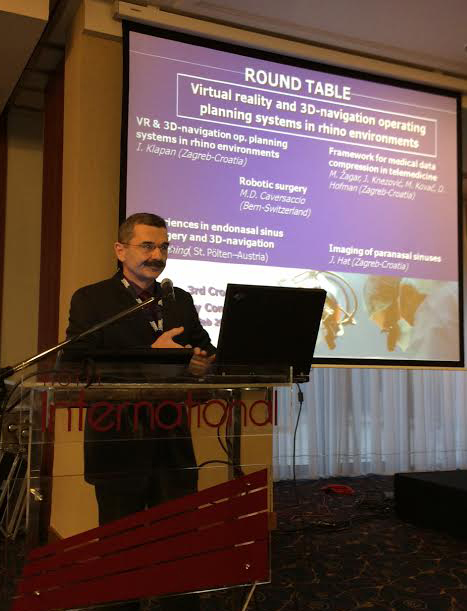
I. IV. 3. CROATIAN RHINOLOGIC CONGRESS, ZAGREB, HRVATSKA (Veljača 2014.) http://www.3rhinocongress.org/programme.asp
1. OKRUGLI STOL (1st ROUND TABLE) (Prof. Klapan/voditelj/Chairperson): CAS - Virtual reality and 3D-navigation operating planning systems in rhino environments
Invited speakers:
I. Klapan (CRO), M.D. Caversaccio (CH), T. Fasching (A),
M. Žagar (CRO), J. Hat (CRO)
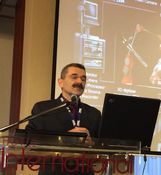
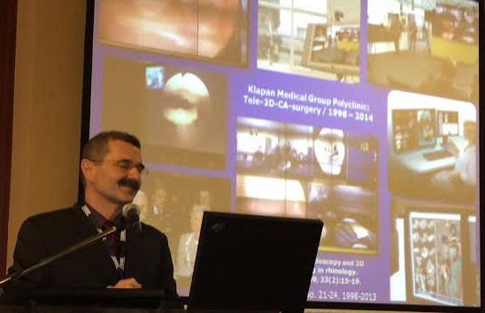
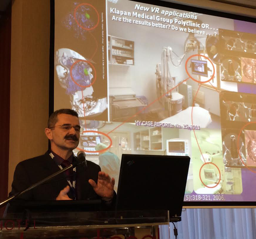
Additional research in the area of 3D image analysis, visualization, tissue modelling, and human-machine interfaces provides scientific expertise necessary for developing successful 3D visualization of the human head during 3D-CAS, Tele-3D-CAS, and other VR (virtual reality) applications. Such an impression of immersion can be realized in any medical institution using advanced computers and computer networks that are required for interaction between a person and a remote environment, with the goal of realizing tele-presence. If we would like to understand the idea of VR, it is necessary to recognize that the perception of surrounding world created in our brain is based on information coming from the human senses and with the help of a knowledge that is stored in our brain. The usual definition says that the impression of being present in a virtual environment, such as virtual/tele virtual endoscopy of the patient’s head, that does not exist in reality, is called VR.Now, imagine that we can substitute artificially generated sensations for the real standard daily information received by our senses. In this case, the perception system in humans could be deceived, creating an impression of another 'external world around the man (e.g., during standard 3D navigation surgery). In this way, we could replace the true reality with the simulated reality that enables precise, safer and faster diagnosis as well as surgery. All systems of simulated reality share the ability to offer the user to move and act within the apparent worlds instead of the real world. Any otorhinolaryngologist, member of my surgical team, has impression of presence in the virtual world and can navigate through it and manipulate virtual objects (www.poliklinika-klapan.com). We are introducing guidelines and suggestions for use of 3D image processing SW in head pathology diagnostic and procedures for obtaining physical medical model by additive manufacturing/rapid prototyping techniques, bearing in mind the improvement of surgery performance, its maximum security and faster postoperative recovery of patients. This approach has been verified in our numerous case reports. In the treatment we used intelligent classifier-schemes for abnormal patterns using computer-based system for 3D-virtual and endoscopic assistance in rhinology, with appropriate visualization of anatomy and pathology within the nose, paranasal sinuses, and scull base area. In human medicine, once created from 2D-CT/MRI cross-section images with 3D modelling SW, virtual 3D surface models or VR models can be further used in VR for measuring, operation planning, simulations and structural finite element analysis (FEA). Virtual 3D models also can be used in actual reality for tangible, physical (real) models obtained e.g.by rapid prototyping (RP) technique. RP techniques look most promising to satisfy medical need for tangible models. While prototyping is a usually slow and expensive process of building pre-production models of a product to test various aspects of its design, RP techniques are methods that allow quick production of physical prototypes. Once created from 2D cross-section images with 3D modelling software, virtual 3D surface models or volume rendered models can be further used in virtual reality(VR).Our final experience in the field of use appropriate virtual medical models, indicate that the entire diagnostic and/or operative procedure can be completely simulated, and critical areas avoided during the real procedure by employing real patient images in the operation preparatory phase using complex spatial models and simulated operative field entry (RP/rapid prototyping models, VE/virtual endoscopy, VS/virtual surgery). Systematic approach combined with modern tools for medical imaging and additive manufacturing provides: computer-based system for virtual endoscopic assistance in rhinology, extending physician’s visualization and orientation in the anatomical space of the nose and surrounding areas, comprehensive detailed 3D procedure planning with possibility to prepare and consider several scenarios quickly, follow-on illustration-piloted endoscopy, real-time processing and fusion of the 3D MSCT data and endoscopic video that helps preventing undesirable intrusions into sensitive areas with instruments during surgical procedures, additional visual tools about objects examined/implemented in system, like layers, volume, surface and distances, tangible and realistic models produced with additive manufacturing technologies, the physical models of human organs those are extremely effective in realizing the anatomy, virtual planning and simulation of operation various surgeries, with complex spatial relationships. ABSTRACT CODE: 327ZUUA126R8Z7A
References: Klapan I, et al. (2006) Ear Nose Throat J, 85(5):318-321; (2008) Coll Antropol, 32(1):217-219; (2011) Virtual Reality in Medicine, ISBN 978-953-307-518-1, Intech, 303-336.
.jpg)